Composition of Semen, Male Reproductive Anatomy, and Forensic Importance — A Complete Guide
Semen is a biologically complex fluid essential for human reproduction and a key biological evidence type in forensic investigations. This guide explains semen composition, sperm formation (spermatogenesis), male reproductive anatomy, the secretions of accessory glands, comparative semen volumes across species, and why semen matters in forensic contexts.
1. Introduction to Human Reproduction
Human reproduction depends on the fusion of two specialized sex cells: sperm (male gametes) and the oocyte (female egg). When a sperm fertilizes an oocyte, the resulting zygote develops into an embryo. The male gamete's journey from production to fertilization is supported and enabled by seminal fluid—commonly called semen.
2. What Is Semen?
Semen is a liquid or semi-gelatinous cellular suspension that contains spermatozoa and secretions from the male accessory reproductive glands. Sperm do not travel alone; they are mixed with a complex cocktail of fluids that protect, nourish, and enable sperm motility and viability within the female reproductive tract.
3. Male Reproductive Anatomy — Overview
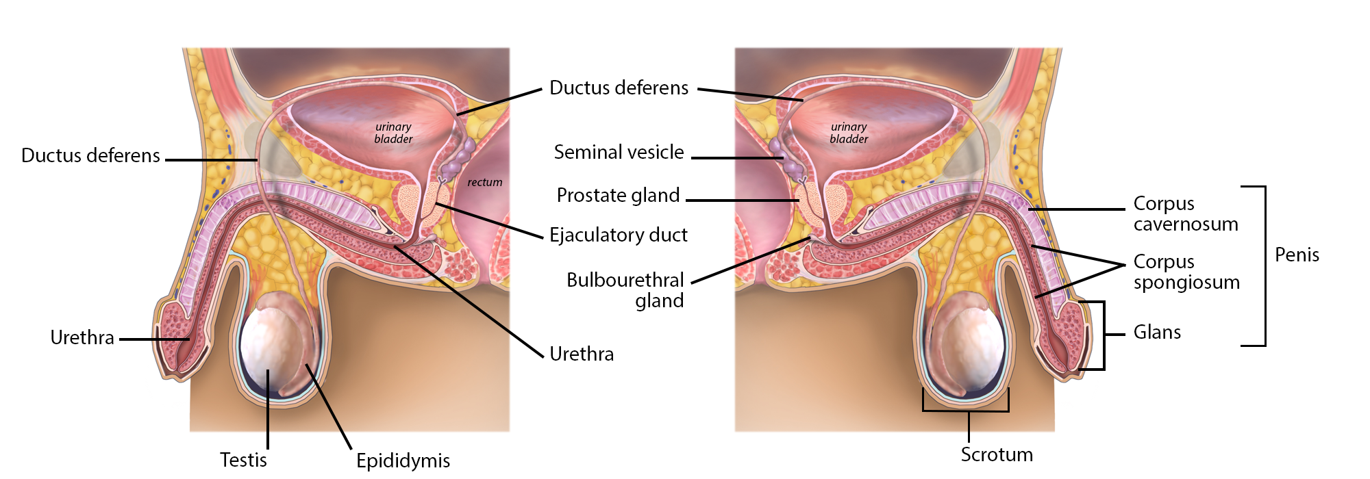
The male reproductive system consists of the testes, a series of ducts (epididymis, vas deferens, ejaculatory duct, urethra), and several accessory glands (seminal vesicles, prostate gland, bulbourethral glands). Together they produce, mature, transport, and expel sperm and seminal fluid.
3.1 Pathway of Sperm
- Testes (sperm production)
- Epididymis (maturation & storage)
- Vas deferens (transport)
- Seminal vesicles (major secretions)
- Ejaculatory duct & prostate (mixing)
- Urethra (expulsion)
4. Testes and Spermatogenesis
4.1 Testes — structure & function
Each testis is an ovoid organ (≈ 5 cm long, 3 cm wide), located in the scrotum. The scrotum keeps testicular temperature about 3°C lower than core body temperature—an essential condition for efficient sperm production.
4.2 Seminiferous tubules & histology
Testes contain many coiled seminiferous tubules, where spermatogenesis occurs. Supporting cells include:
- Sertoli cells — nourish developing sperm and maintain the blood-testis barrier.
- Leydig cells — interstitial cells that produce testosterone.
4.3 Spermatogenesis (step-by-step)
Sperm develop through progressive stages within the seminiferous tubules:
- Spermatogonia (stem cells)
- Primary spermatocytes
- Secondary spermatocytes
- Spermatids
- Spermatozoa (mature sperm)
The complete maturation process takes roughly 2–3 months. The slides depict these stages and the morphological transformations (head, midpiece with mitochondria, and flagellum) required for motility and fertilization.
5. Epididymis, Vas Deferens & Ejaculatory System
Epididymis
The epididymis is a highly coiled duct (about 18 feet when stretched) that sits along the testis. Sperm spend about 20 days here to gain motility and fertilizing capability; older sperm are reabsorbed.
Vas deferens & ejaculatory duct
The vas deferens transports sperm from the epididymis toward the pelvic cavity; it unites with seminal vesicle ducts to form the ejaculatory duct, which passes through the prostate and empties into the urethra.
Urethra
The urethra is the final passage for semen. During ejaculation, semen is expelled through the urethral opening at the penis.
6. Accessory Glands & Seminal Fluid Composition
Most of semen’s volume and biochemical properties come from accessory glands. Their secretions together create an optimal medium for sperm survival and function.
6.1 Seminal Vesicles (major contribution)
Seminal vesicles are paired glands that contribute roughly 70% of ejaculate volume. Their secretion contains:
- Alkaline fluid — neutralizes acidity of male urethra and female tract.
- Fructose — a key energy source for sperm motility.
- Prostaglandins — modulate immune response and may aid sperm transport.
- Clotting factors — help semen temporarily coagulate post-ejaculation.
6.2 Prostate gland
The prostate adds an enzyme-rich, milky fluid that helps activate sperm and contributes to initial ejaculatory fraction.
6.3 Bulbourethral (Cowper’s) glands
These paired glands secrete clear mucous that lubricates the urethra and neutralizes residual acidity prior to ejaculation.
7. Semen Volume Across Species (comparison)
Semen volume varies greatly between species—information relevant to veterinary reproduction and comparative studies. Typical human ejaculate is approximately 2–6 mL.
| Species | Typical Volume (mL) |
|---|---|
| Man (Human) | 2–6 |
| Boar | 150–500 |
| Stallion | 30–300 |
| Bull | 2–10 |
| Dog | 2–15 |
| Ram | 0.7–2 |
| Rabbit | 0.4–0.6 |
8. Forensic Importance of Semen Examination
Forensic science leverages semen analysis for multiple investigative purposes:
- Detection in sexual assault cases: Identification of spermatozoa on swabs or clothing, and biochemical tests (e.g., acid phosphatase, PSA) to confirm seminal fluid.
- DNA profiling: Sperm cells and other cellular material in semen provide high-quality DNA for individual identification and linkage to suspects.
- Estimating time since intercourse: Patterns of sperm decay and persistence in different body sites assist timeline reconstruction.
- Differentiation from other fluids: Biochemical and microscopic methods help distinguish semen from saliva, urine, or vaginal secretions.
9. Practical Takeaways
- Multifunctional fluid: Semen supports sperm physically, biochemically, and immunologically during the fertilization process.
- Accessory glands matter: Seminal vesicles, prostate, and bulbourethral glands each contribute distinct components essential to semen function.
- Forensic relevance: Semen detection and DNA profiling are cornerstone techniques in sexual assault response and criminal investigations.